High Arched Foot
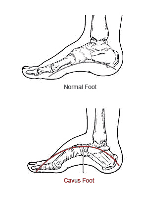
The cavus, or high arched foot, is described as having a high medial longitudinal arch. A patient may have a reducible or rigid cavus foot structure. There are many etiologies for a cavus foot type. Many stem from an inherited neuromuscular disorder. Not all high arched feet are symptomatic. In some, symptoms may occur in the foot, ankle, or leg. Claw toes are very common with cavus foot types. Painful callosities may develop on the dorsal aspects of the digits and the plantar aspects of the metatarsal heads. With hindfoot or combined cavus, a prominent posterior aspect of the calcaneus may occur, which can be irritated by the heel counter of the shoes. Severe cramps in the legs may result from a true or pseudo Achilles tendon equinus. In conditions caused by peripheral nerve pathology (i.e., Charcot Marie Tooth disease) tendon imbalances may occur, including the loss of entire muscle groups which can lead to a drop foot condition.
Proper diagnosis is needed to assess whether neurologic pathology is present and, if so, to determine its classification and stage. This is important to determine if the condition may progress overtime.
The cavus (high arch) is a clinical challenge, especially in progressive conditions. Many times conservative treatments provide pain-free ambulation. Treatment is initially aimed at alleviating painful callosities by proper shoeing, accommodative orthoses, or an AFO (ankle foot orthoses) if a drop foot is present. If conservative and palliative measures fail to alleviate symptoms, surgical reconstruction is considered. Using a combination of soft tissue(tendon transfers, releases) and osseous procedures, the painful cavus foot is reconstructed. For some cavus conditions, digital deformities (single or multiple) are all that need correction, but for others, multiple hindfoot fusions with tendon transfers from the posterior leg are necessary.
